NucMedResource.com
  • Home
  • Radiology
  • Blog
  • Favorite Books
  • Pulmonary NM ("V/Q")
  • Endocrinology NM
    • Benign Thyroid Disease
    • Thyroid Cancer
    • Parathyroid Scan
  • Neurology NM
    • Brain Death Scan
    • DaT Scan
  • Neurology PET
  • Oncology NM
    • MIBG
    • OctreoScan
    • ProstaScint
  • Oncology PET
    • Normal FDG PET Oncology
    • PET Lung Cancer
    • FDG PET Pitfalls
  • Cardiology NM
    • EKG
    • MP SPECT
  • Cardiology PET
  • GI
    • Hepatobiliary "HIDA" Scan
    • Gastric Emptying
  • Bones
  • Infection & Inflammation
    • WBC Scan
    • Gallium Scan
  • Other General NM
  • Lymph Node Stations
    • H&N Nodal Stations
    • Thoracic Nodal Stations
    • Abdominal Nodal Stations
  • Protocols
  • Presentations
  • Reading Material
  • Exposures
Back to Staging Cancers

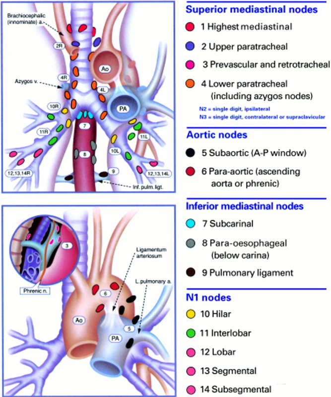
Classifying Thoracic (Mediastinal & Hilar) Lymph Nodes

For many cancers and especially lung cancers, it is important to describe the location of an abnormal lymph node in a way that clinicians who may biopsy these nodes can understand. These figures offer guidance on the standardized way to describe these nodal stations. 
Picture
Source
Fountain SW. Guidelines on the selection of patients with lung cancer for surgery. Thorax 2001;56:89-108.

Helpful
Smithuis R. "Lymph Node Map." The Radiology Assistant. 
This site is educational in nature and for nuclear medicine, radiology and medical professionals in the U.S., contains user contributed content, and material that may be confusing to a lay audience. Readers are asked to check the most current information on nuclear medicine techniques as this website may not be all-encompassing or up-to-date. It is solely the responsibility of the professional to practice nuclear medicine based on his or her experience and knowledge of the patient. To the fullest extent of the law, neither the publisher or authors assume any liability for any consequences to persons or property arising out of or related to any use of the material in this website.