Nuclear Cardiac Imaging
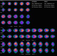
The most common study in this category is myocardial perfusion SPECT imaging ("MPI," erroneously referred to as "P. thal") for suspected coronary artery disease, but nuclear imaging is also used to evaluate LV ejection fraction ("MUGA").
Reviews on Myocardial Perfusion SPECT
Burrell S and MacDonald A. Artifacts and Pitfalls in Myocardial Perfusion Imaging. J Nucl Med Technol 2006;34:193-211.
Dvorak RA et al. Interpretation of SPECT/CT Myocardial Perfusion Images: Common Artifacts and Quality Control Techniques. RadioGraphics 2011;31(7).
Dvorak RA et al. Interpretation of SPECT/CT Myocardial Perfusion Images: Common Artifacts and Quality Control Techniques. RadioGraphics 2011;31(7).
Protocols
Procedure Guideline for Myocardial Perfusion Imaging (ver 3.3, 2008)
Appropriateness Criteria
Basic EKG Information
As understanding of basic EKG findings is important for proper interpretation of Myocardial Perfusion Imaging. Click here to read more.
Backlog
SPECT imaging for detecting coronary artery disease and determining prognosis by noninvasive assessment of myocardial perfusion and myocardial viability. (PubMed link)
Cardiac PET/CT for the Evaluation of Known or Suspected Coronary Artery Disease. (PubMed link)
Interpretation of SPECT/ CT Myocardial Perfusion Images: Common Artifacts and Quality Control Techniques. (PubMed link)
Cardiac PET/CT for the Evaluation of Known or Suspected Coronary Artery Disease. (PubMed link)
Interpretation of SPECT/ CT Myocardial Perfusion Images: Common Artifacts and Quality Control Techniques. (PubMed link)